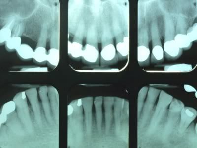
Why are Digital Dental X-rays Better Than Film?

I came across answer.yahoo and found out some answers related to this question, "Why are Digital Dental X-rays Better Than Film?" Here are some of the inputs from professional dentists and registered dental hygienist. Check this:

----------------------
X-rays allow us to see the spaces in between teeth, underneath crowns and fillings, and to check the bone levels underneath the gum line. Occasionally a specific tooth needs to be investigated for decay or infections with another X-ray. Our clinicians will determine how often your X-rays need to be taken.

The digital dental X-ray system is more sensitive than conventional X-ray film systems, so your exposure to X-rays is cut by as much as 90 percent. The large, colour-enhanced image is displayed on a computer which gives both our clinicians and patients a clearer picture of teeth, bone and surrounding oral structures.

Digital X-rays have less of an impact on the environment. There are no used photo chemicals or lead films to dispose of. Because there's no time wasted waiting for films to be developed, your appointments take less time and it's fun to watch this system work! Most of our patients are amazed at how quick and easy the process is.

With better resolution, dramatically reduced radiation to the patient and the ability to zoom into parts of the image, digital dental X-ray is friendlier to our patients, and to the clinician. In addition to this, digital X-rays provide the ability to archive in original excellent quality, meaning that we can compare current X-rays to previous X-rays, and to send a perfect digital copy to specialists if the need arises.

Source: Greg Dougall Dental gdougalldental.com.au

----------------------

You get less radiation and they are able to see the film more clearly. Because it's linked to a computer system they can change the contrast, measure bone level, clarify the image, diagnose more accurately, the list is endless. Digital x-rays are much better for you and them. You should be proud that your office is technologically advancing

Source: RDH, B.S (Registered Dental Hygienist

----------------------

Digital xrays will expose the patient to less radiation and will allow the person taking the xrays to see them immediatly.
Source: Ronswife (Certified dental assistant)

----------------------

There are five major reasons (in order of importance)
1.) *Less radiation exposure for the patient*,
2.) Better images for diagnosing,
3.) Earth friendly: no more hazardous chemicals generated during processing.
4.) Faster imaging,
5.) Real time worldwide electronic portability and accessibility with no degradation of image.
Glad your dentist made this commitment. It's a big deal to change over the office. Obviously s/he feels patient safety and health is important.
Source: Rayen | Another digital dentist

Source: answers.yahoo.com


Why Most of the Dentist in Bangalore are asking to adopt Digital X-rays?

Here's a news I got from dnaindia.com. One of the dentist in Bangalore said “One such practice is that we are asking them to adopt digital X-rays. Many chemicals are used in developing traditional X-rays, and even the plastic sheets on which those X-rays are printed, are non-degradable. Digital X-rays can be transferred to a computer and later to a CD for future use. They can also be e-mailed,”

Read the complete story below:


Dentists vow to go green to keep your teeth white

Dentists in Bangalore city have vowed to go green. They have started a go-green campaign to make dental clinics eco-friendly.

“We are aiming to promote the concept of green dentistry. There are many practices in this profession that need to be changed to achieve this goal. The theme of the campaign is ‘good for you and for the earth’,” said Dr Asish Shetty, professor, Bangalore Institute of Dental Sciences and Hospital. He was talking on the sidelines of Rotary Marathon 2010, a workshop organised by the Bangalore Dental Association on Tuesday. The green dentist concept will touch all fields of the profession —from conserving water and electricity to avoiding use of chemicals that are harmful to the environment.

“The amount of water wasted by a person while brushing will come up to 90 glasses, approximately. We need to bring down such losses. If we replace spotlights we use in clinics with CLF bulbs we can save lots of energy. We also need to stop practices like silver filling of root canals. The chemicals used in silver filling are also damaging to the environment. We are using the technology as the alternatives are highly expensive. If we can do without such practices, it will do lots of good for humanity,” he said.

Dentists across the city are being made aware of the need to adopt eco-friendly methods as part of the campaign. “One such practice is that we are asking them to adopt digital X-rays. Many chemicals are used in developing traditional X-rays, and even the plastic sheets on which those X-rays are printed, are non-degradable. Digital X-rays can be transferred to a computer and later to a CD for future use. They can also be e-mailed,” said Swaria Rao, director, Bangalore Institute of Dental Sciences and Hospital.

Dentists participating in the go-green campaign will be given badges. “The concept behind the badges is that they will help promote the idea even among the patients. When they go to hospitals wearing the badges, patient will enquire about them, which will further spread our message,” said Dr Rao.

“Patients also play a vital role in such ventures. A toothbrush which can be used for four months is thrown away by people in just three weeks. Such practices should also be changed,” she added.

Published: Wednesday, Sep 15, 2010, 10:01 IST |By Arun Dev | Place: Bangalore | Agency: DNA

source: http://www.dnaindia.com


Digital X-Ray Technology - An Owner's Perspective

By Terry L. Myers, DDS, FAGD

In 2006, 735 Dental Economics® subscribers were polled about various aspects of dental practice, including their aspirations for new technology. Dentists rated radiography equipment as the highest priority in the basic equipment investment technology category. At the time, 29% of the respondents were planning to add a digital system in the next two years, and a surprising 41% responded that they had already implemented a digital radiography system.

Since that time, other surveys have echoed these statistics, with technology taking an even more important role in the dental practice. Additionally, research and development in digital radiography continues to make this a technology that counts both clinically and financially. In the past, I have been an outspoken advocate for imaging, but with so many dentists yet to adopt the digital-office lifestyle, it's worth sharing again.

While every office has its own priorities, these are the top three benefits of digital X-ray technology that have enriched life around the office for me, my staff, and my patients.

More than meets the eye » Digital X-rays are as clear and detailed as any conventional film, and the digital format offers choices. There's really not much chance for education to occur with a 1" by 1½" piece of film. However, education with digital X-rays is phenomenal. Images are the size of the computer monitor. If I want to see different perspectives, I can magnify, zoom in, change color and contrast, draw on, and point out my concerns with a mouse click until I am absolutely sure that the patient sees the situation from my viewpoint and is confident in my assessment. Innovations in digital technology just keep getting better. Images taken with the DEXIS Platinum sensor that I recently obtained combine all sorts of technology to bring out clarity in even hard-to-see details.

More time for patients » Digital X-rays are faster than film. For example, my system can capture vertical or horizontal bitewings and periapicals with a single sensor – without changing film or sensors. My assistant can take an FMX in about five minutes. When my assistants are on the move, the sensor moves with them; the direct USB connection allows quick and easy transport between treatment rooms. With digital X-rays literally at my fingertips from any office computer or even from home, I can securely pull up images and records for an informed decision. Try doing that with film.

What's best for diagnosis and patient education?


It's just more practical » With all the attributes of digital radiography, clinicians often forget the more practical side of producing X-rays – the equipment that produces the X-rays. The processor is a high-maintenance piece of equipment. Mechanical parts break down or start slipping, and then it has to be sent out for repair, so you have to get a spare or a loaner. Besides, it is not practical for assistants to spend time developing film or cleaning the processor when they could be caring for patients.

While these are my top three benefits, digital radiography also brings a sense of cohesiveness to all of the office processes. When integrated with a digital practice-management system, digital image files can be stored with treatment notes and records, appointments, and insurance information – all in one organized, uncluttered space.

While technology can touch every aspect of the office these days, keep in mind that the X-ray is the first image that patients see, and as such can establish your image as a technology-savvy dentist.

For more on this topic, go to www.dentaleconomics.com and search using the following key words: dental technology, digital imaging, X-ray, dental radiography, diagnosis, Dr. Terry L. Myers.

Dr. Terry L. Myers is a fellow in the Academy of General Dentistry and a member of the Academy of Cosmetic Dentistry and the Dental Sleep Disorder Society. He has a private practice in Belton, Mo.


Source: http://www.dentaleconomics.com

Introducing the Gendex GXCB-500 Cone Beam 3-D Imaging System

Another innovation of Cone Beam 3D Imaging System. Introducing the Gendex Dental Systems: GXCB-500 Cone Beam 3-D Imaging System.


Video source: http://www.dentalcompare.com/video_view.asp?videoid=242


Video Source: http://dentalcompare.com/details/9106/GXCB-500-Cone-Beam-3-D-Imaging-System.html

General Information
VendorGendex Dental Systems
ItemGXCB-500 Cone Beam 3-D Imaging System
Product NumberGXCB500
WarrantyInquire
Focal Spot0.5
Footprint48 inches wide x 46 inches deep
Scan Time8.9 sec (.3 and .4 voxel, Standard/EDS Mode)
23 sec (.125, .2 and .25 voxel, Standard Mode)
Installation IncludedYes
Patient PositioningSeated
Primary Reconstruction Time<>
Volume ReconstructionYes
Panoramic ReconstructionYes
Cephalo ReconstructionInquire
DICOM ExportYes (DICOM 3 Compatible)
Plate Dimensions13 x 13 cm
Image DetectorAmorphous Silicon Flat Panel
Software OSInquire
Included Softwarei-CAT Vision™
Gray Scale14 bit
Voxel Size (Slice Thickness).4, .3, .25, .2, .125
Other Included ItemsTraditional Pan imaging plus Cone Beam 3-D
Additional OptionsInquire
Training IncludedInquire

Gendex offers the GXCB-500, an affordable Cone Beam 3-D imaging system powered by industry-leading i-CAT technology. Unique to the GXCB-500 is a ‘medium’ scan area of one or both arches that can extend to encompass the TMJ area, making the system a powerful diagnostic and treatment-planning tool for implant planning, TMJ analysis, and a variety of surgical procedures. The GXCB-500’s Amorphous Silicon Flat Panel sensor yields accurate, highly detailed images at one of the fastest scan and reconstruction rates in the industry. Adding to its value, the system also serves as a traditional 2-D digital panoramic without changing sensors.

Source: HERE

Digital Dental Radiography Review: The DEXIS Digital Diagnostic System

Here's an article by Andrew W. Chin, DDS about digital radiography.

The DEXIS Digital Diagnostic System
By Andrew W. Chin, DDS

Digital radiography is not new to dentistry. It has been in use for over 10 years, and is only becoming more popular and mainstream. There are many companies to choose from. I believe that the DEXIS system is one of the better digital radiography systems on the market today. In general, all digital radiographic systems will reduce the radiation exposure to your patients by about 30-40%. They also reduce the chemicals and raw material waste that go along with traditional radiographs. The quality of digital radiography was always the question, as with traditional photography and digital photography. However, as with digital photographs, digital radiography is getting better (if not surpassing) the traditional films, as our technology improves.

The DEXIS Digital Diagnostic system has been on the market since nearly the beginning of digital radiography. In those years it became the first company to have its digital radiography system receive the American Dental Association Seal of Approval when it was admitted into the ADA Acceptance Program. Recently, DEXIS received the Reality Magazines five-star award for outstanding quality in a digital radiography system. These awards only confirm what I have known through the years working with the system.

The DEXIS hardware is very unique. Most other systems have multiple-sized sensors. The DEXIS system has only one size and the corners are rounded for comfort. Although it is one of the thicker sensors on the market, the rounded corners, which other systems do not have, make a significant difference in patient comfort. Many gaggers have even said that the sensors are much more comfortable and create less of a gag reflex. The sensor and its hardware are easily transferred to adjacent rooms and computers. If your operatories are not networked, the unit can be used on a laptop. The sensor is the weakness of all digital radiograph systems due to its mobility and small fragile size. DEXIS is no different in this respect.

The sensor costs around $6,000 to replace, and about half that if you buy the insurance program and software support. This will run you a yearly fee of about $1,300-$1,800. You can weigh the costs and benefits of not buying the maintenance program. Regardless of what you choose, your assistants will need to treat the sensor with great care.

The entire office will go through a training program provided by the company. The learning curve is fairly fast and not too complex, especially if you are familiar with computers. The DEXIS radiographic system has some excellent clinical features that will improve your clinical diagnosis and efficiency. The actual radiograph will be instantaneous after the radiation has been detected. This image can then be enlarged to fill your monitor, magnified at specific areas, or the resolution can be adjusted at the click of a mouse. There are many other bells and whistles that I do not use, but the most useful feature is something they called ClearVu. This feature will greatly enhance your clinical diagnosis and be more accurate in finding decay or other pathology. Lastly, the DEXIS system has very convenient measuring tools for your endodontic and implant procedures. With so many features, the DEXIS system will help educate your patients and you will see your treatment plans increase in acceptance.

Cost will always be a factor in these types of high tech equipment. The entire system will run over $10,000, and don’t forget those yearly maintenance costs. The DEXIS system will have other costs in bridging software if you want/need to integrate it with your practice management software. I have integrated it with mine and have had no complications. Even with these costs the system will save you time and will make you much more efficient. Ultimately you will produce more because of the increased efficiency and the ease of presenting your treatment plan by educating patients with the DEXIS system.

DEXIS is the most highly awarded digital X-ray system, well-known for unique features like "One-Click FMS” and ClearVU™ image enhancement. The new DEXIS Platinum Sensor with PureImage™ technology has three distinct advantages:
  1. A smart design that places the electronics on the sensor’s back so the active imaging area is almost 100% of the total sensor size.
  2. An advanced ergonomic design featuring four rounded corners, a slim profile, and a seamless casing, allowing precise sensor placement while assuring optimal patient comfort.
  3. A patented cord placement that allows for vertical AND horizontal bitewings and all peripherals with a single sensor, thus eliminating the cost and inconvenience of multiple sensors of different sizes.
DEXIS features a direct USB connection, with no controllers or docking stations. DEXIS also offers easy integration with practice management programs, digital pan systems, cameras, and scanners allowing the clinician to capture, store, and access all X-ray and camera images through one convenient Imaging Hub.

For more information about DEXIS product check here

Article Source: http://www.dentalcompare.com/review.asp?rid=10

Dental Imaging Software Application

Dental imaging software offers easy-to-use, full-featured dental programs that completely integrate dental practice management, digital dental imaging and patient clinical charting.

For more complex imaging requirements, dental practitioners really need dental imaging software. It is very common to use one program for dental practice management and a different program for image management. If you are using digital radiography, then your hardware vendor will have an imaging program you can purchase. Examples include Dexis, Schick, and Gendex VixWin. There are also independent software products such as XDR, TigerView, and Apteryx which work very well.,

ADSTRA is one of the company promoting dental imaging software solution. ADSTRA Imaging™ is a world class, comprehensive software designed to manage dental images provides easy image capture from intraoral cameras, video microscopes, scanners, and intraoral, panoramic and cephalometric digital x-ray devices. With ease and flexibility, ADSTRA Imaging™ enables users to view images when they want them.

Another company who offers dental imaging softwre is Kodak. They have dental imaging software for Windows. KODAK Dental Imaging Software serves as the control panel for all of our digital imaging systems. Its flexible user interface has been designed specifically for dental radiological diagnosis, and can be used as a standalone program or integrated with your practice management software.

There are other companies providing dental imaging software that you can choose from. These dental imaging software will always depend on your dental imaging equipment.

Alarming News: Ten Dental X-rays 'Raise Cancer Risk'

Dental X-rays given to millions of Britons every year may dramatically increase the risk of thyroid cancer, scientists warned last night.

Researchers found that patients who had been X-rayed by their dentist at least ten times were more likely to develop the disease.

They have now warned that X-rays should not be given at check-ups or when registering new patients - despite these practices being common in many dental surgeries.


Regular dental check-ups are important to maintain healthy gums and teeth, but scientists have found a link between dental x-rays and thyroid cancer

How the dental X-rays work when a patient visits the dentist for a check up

With rates of thyroid cancer more than doubling in 30 years, the scientists said that the potential dangers of dental X-rays were often overlooked.

Researcher Dr Anjum Memon, of Brighton and Sussex Medical School, said: 'Our study highlights the concern that, like chest or other upper body Xrays, dental X-rays should be prescribed when the patient has a specific clinical need, and not as part of routine check-up or when registering with a dentist.'

'A Safe and valuable tool in caring for our patients' By Dr Martin Fallowfield Peterborough dentist and British Dental Association spokesman

X-rays are a vital tool across medicine and dentistry.

Dentists employ them in one of two ways: either as a screening device as part of a thorough examination to assess gums, teeth or bones, or to help investigate a particular problem or prepare for a procedure such as implants.

X-rays can reveal problems that simply can't be observed by the naked eye. Tooth decay, for instance, can be far more extensive than it appears.

An X-ray provides the full picture - often before pain or discomfort make the full extent of the problem plain to the patient.

The general principle dentists adhere to is that as few dental X-rays as is practicably possible should be taken. But each patient is different, and their care requires dental X-rays at different intervals and for different reasons.

The need for X-rays won't go away though, and dentists are required to undertake training to ensure they are up to date with the latest techniques in deciding when and how to take X-rays.

That keeps this hugely valuable tool safe and at our disposal in caring for our patients.

Dental leaders recommended that patients protect their thyroid - a hormone-releasing gland at the base of the neck - by wearing lightweight lead collars or bibs when being X-rayed.

The researchers asked 313 thyroid cancer patients and a similar number of healthy volunteers how many dental Xrays they had undergone.

After factoring in any hospital X-rays participants had had, they found that men and women who had had up to four dental X-rays were more than twice as likely to have developed the disease than those who had never had any.

Between five and nine X-rays and their risk rose more than four-fold, the journal Acta Oncologica reports.

In most danger were those who had had ten or more X-rays - their risk was 5.4 times that of someone who had never been X-rayed in the dentist's chair.

The researchers relied on patients' recollections, rather than dental records, but said that despite this, the finding was significant.

Dr Memon added that the results were supported by previous reports of increased risk of thyroid cancer in dentists, dental assistants and X-ray workers, suggesting that multiple low-dose exposures may be harmful.

Dr Memon, who carried out the research with experts from Cambridge and Kuwait universities, said: 'It is important that our study is repeated with information from dental records including frequency of X-rays, age and dose at exposure.

'If the results are confirmed, then the use of X-rays as a necessary part of evaluation for new patients, and routine periodic dental radiography, at six to 12 months interval, particularly for children and adolescents, will need to be reconsidered, as will a greater use of lead collar protection.'

But British dentists pointed out that the study was carried out in Kuwait, where rates of thyroid cancer are much higher than in the UK, and said the researchers did not know what sort of X-ray equipment had been used.

Dr Nigel Carter, chief executive of the British Dental Health Foundation, said: 'The number of X-rays being taken in dental practice in the UK has greatly reduced in recent years and the dosages from modern equipment are extremely low.

'Dental X-rays have a valuable role in the diagnosis of dental disease.'

Source

Dental Imaging Review: Progeny Dental JB-70 Dental Intraoral X-Ray

The JB-70 redefines AC technology with the perfect balance of imaging excellence, simple, efficient operation and smart good looks. Microprocessor controls and our advanced pre-heat feature make the JB-70 the imaging foundation of today’s dental practice—ideal for standard and high speed films and optimized for today’s digital receptors. It’s a new generation of dental imaging brought to life in an intraoral x-ray system that is smarter, more reliable and easier to use.

All the features you need for everyday imaging excellence:
  • 70 kVp and 7 mA
  • 0.7 mm focal spot
  • Large, easy to understand icons
  • 30 Pre-programmed techniques
  • Manually selectable time settings
  • Effortless, drift-free positioning
  • Unique tube head handle
  • Compatible with film, sensors and phosphor plates


Intuitive and Efficient Operation

Easy to understand icons and 30 pre-programmed anatomical technique settings ensure instant proficiency for every user. Time settings can be selected according to individual preferences using the keys on the control panel and stored into memory for future use. The sleek control panel can be removed from the power base and mounted in another area in or outside of the operatory. Two push button remote stations and a corded control are also available as accessories for remote exposure installations.

Sophisticated materials and design provide unprecedented quality and stability

High-tolerance, frictionless joints provide amazingly smooth and effortless arm movement. Add Progeny’s heavy-duty, dual braking system for dependably precise and stable positioning for years to come. The JB-70 is available with a 56”, 66” or 76” arm length and can be mounted on the wall or in a pass through. The JB-70’s tube head handle makes movement especially easy and the offset tube head design allows the user to see the patient clearly at all times for precise positioning.

Here's a product review I got from this site: http://dentalinsider.wordpress.com

Progeny JB-70 X-Ray Product Review

What to do get when you take Engineers, Vice Presidents and the CEO of one of the leading companies in dental xray and give them a clean slate to start from?
Progeny Dental’s JB-70….The Next Generation of Dental Imaging has arrived. This xray not only looks good, but offers simple controls, flawless imaging and a very competetive price. ($4500.00 Retail)

Intallation is very straight forward and is setup for a single stud mounting, right out of the box. An optional two stud mounting plate($185.00 retail) is available when you’re mounting the unit across two studs, 16 inches on center.

If you’re in the market for a new x ray unit, I suggest you take a look at the Progeny JB-70 or their sister xray unit the Preva DC unit. Either would be a smart choice for your practice.


Source: progenydental

For complete details check here

Dental Digital Radiography News: Vetel Diagnostics Introduces Envision Dental™ Diagnostic Imaging Tools

PRLog (Press Release) – Jul 27, 2010 – Vetel Diagnostics, a leading provider of diagnostic imaging tools to veterinarians, introduces ENVISION DENTAL™, a new line of dental digital radiography (DR) products: the AnyRay handheld dental DR generator, the EZ Sensor line of dental DR detectors, and the new Metron dental software featuring an interactive positioning guide. They are designed to make dental imaging easier and to increase the veterinarian’s opportunities to deliver dental services.

An estimated 80% of canine and feline populations have some type of treatable dental pathology. Despite the prevalence of periodontal disease, however, many practices face challenges in delivering even routine dental services. The time and expertise required using traditional radiography presents a significant obstacle during the average appointment. Recognizing the disadvantages this creates for both patient care and practice profitability, Vetel Diagnostic’s pursued imaging solutions to address both.

Dr. James K. Waldsmith, President of Vetel explained: “These tools represent a huge leap forward in providing the dental diagnostics that lead to treatment opportunities in daily practice.” As more practices transition to digital imaging, opportunities represented by dental services continue to gain the recognition they deserve. Developing a product line based on superior quality and ease of use is at the core of Vetel Diagnostic’s philosophy. Metron’s ability to integrate dental radiographs alongside other diagnostic images places more complete patient information at the practitioner’s fingertips.

“First and foremost we looked for products that delivered quality imaging”, said Dr. Waldsmith, “but we also understood that positioning tools, ease of use and workflow efficiency have equal importance if the practitioner is to utilize them on a regular basis. Drawing an owner’s attention to their pets’ bad breath begins the conversation but without fast, high quality dental radiographs to provide the diagnoses, client education and dental services are far more difficult to provide. Access to the proper imaging tools substantially increases those diagnostic opportunities. We chose products according to their suitability for superior patient care and practice productivity.”

Vetel Diagnostic’s ENVISION DENTAL™ was created to meet the specific needs of veterinary practice. EZ Sensors were designed with an emphasis on patient comfort; the AnyRay generator affords complete portability and Metron’s dental software provides technician assistance and client education.

Source: PRlog.org

Digital Dental X-Rays - Benefits and Advantages in the Future of Dentistry

This article will explain what are the advantages of Digital Dental X-rays and the Dis-advantages of the traditional dental x-rays.

Digital Dental X-Rays - Benefits in Dentistry

Digital dental x-rays are becoming the standard of care in dentistry. There are many fine systems in use today by a variety of manufacturers. Digital dental radiography was introduced in Germany in 1995 by the Dexis Corporation and into the United States in 1997. All digital radiology systems consists of:
  • A digital sensor instead of film
  • An image capture card (PCMCIA) or processing device.
  • Dental software for image display
  • Enhancement software
  • Digital image storage capability
When compared to dental film x-rays, digital radiology is equivalent (and in some instances better) than conventional dental film. Because some of the software is able to digitally enhance the image we believe is even better than traditional dental x-ray film. Digital radiology most significant advantage is seen in endodontic (root canals) where the dental file and the apex of the tooth need to be visualized in great detail.

Digital dental x-rays reduces radiation exposure to the patient and dental staff.

Digital dental radiography can produce a high quality dental image with 60%-80% less exposure than traditional film dental x-rays. While traditional film x-rays have a very low exposure level ... this takes radiation exposure to a new level (very low).

Digital dental x-rays are friendly to the environment

Traditional dental x-rays use chemicals to develop and fix the image on to the film. These chemicals are not friendly to the environment. In addition, traditional film x-rays use silver halide to develop the image and a silver mixture in not released into the environment. Everyone benefits from this new technology.

Digital x-ray has been around dentistry for some ten years or so. The use of this imaging system benefits all dental patients.

Article Source: James Delapp and H. Candace Delapp (http://EzineArticles.com/?expert=James_Delapp )

How Digital Dental X-Rays Work

Some of the Dentist are now using Digital Dental X-Rays to provide even better dental care and more accurate diagnoses. The process of digitally capturing an image is much faster, safer and more comfortable for the patient.

Sample Digital Dental X-rays Image from the software


How Digital Dental X-Rays Work


Dentist will place a small sensor in your mouth. The sensor is connected to a computer by a thin wire. Next, an X-ray beam is sent through your teeth and into the sensor, which records the image of your teeth and sends it to the computer. The sensor can then be repositioned to photograph other sections of your teeth.


Why Digital Dental X-Rays are Better


The digital dental X-ray system is more sensitive than dental X-ray film systems, so your exposure to X-rays is cut by as much as 90 percent. The large, color-enhanced images let you see what your dentist sees, so it's easier for you to understand how your dentist will treat your teeth. Your fees don't include payment for photo chemicals, film, processing or film storage. Used photo chemicals and film are not polluting the environment. Your dental checkups take less time, and it's fun to watch this system work! Most patients are amazed.

Dental Digital Imaging: Odontogenic Causes of Sinusitis and Cone Beam Diagnostics

By Dr. Mark Weingarden

On May 7th of this year, the University Of Pittsburgh School Of Medicine for Continuing Education in the Health Sciences & Department of Otoloaryngology held their 5th Annual Update in Sino-Nasal Disorders and Allergy. One of the topics discussed was "Clinical Assessment of Dental Pathology for Otolaryngologists and Review of Emerging Dental Imaging".

With the recent addition of the 3D cone beam imaging, a new technology for the dental community, dentists and periodontists have been able to radiographically diagnose periodontal or endodontic problems that may be causing, or contributing to, unresolved or difficult to treat sinusitis in patients.

The trend for the future may be for dental/periodontal practitioners who have cone beam to work hand-in-hand with ENT's and physicians, helping them to be on the lookout for dental issues such as periodontitis and possible endodontic (root canal) issues that may be a possible cause of sinusitis. It is suggested that there may be value in physicians referring their patients for dental or periodontal evaluation, if there is evidence of dental problems. Indications could include:
  1. Red, swollen or bleeding gums
  2. Extensive dental work, crowns, root canals or large fillings in the upper molars
  3. Sensitivity to chewing, temperature or percussion in upper molars
A simple examination to detect potential dental issues can be completed by a physician in 5 minutes, and the instruments needed to do so are few and inexpensive. Some questions that will guide the examiner are:
  1. Is there any tooth discomfort? When did it start? What prompts it? Was any treatment rendered?
  2. When was the patient's last dental exam? How often do they see their dentist?
  3. Does the patient have any crowns, large fillings or root canals in the maxillary posterior teeth?
  4. Is the patient missing upper teeth? When and why were they lost?
  5. Was the patient ever diagnosed with periodontal disease? Do they smoke, have loose teeth or bleeding gums?

Article Source: http://EzineArticles.com/?expert=Dr._Mark_Weingarden

Dental Digital Technology: 21st Century Dentistry - A New World of Dental Technology

Here's an article written by Daniele on how Dental Technology like Dental Imaging and the latest Digital Dental X-rays evolved as an improvement for the 21st Century Dentistry.

21st Century Dentistry - A New World of Dental Technology
by Daniele Johnson

When you meet someone, do you immediately flash him a smile? Or do you hide it because you are in so much pain from tooth ache? Often, people fore go a visit to a dentist despite a tooth ache because of dental fear. A mere sight of the dental chair gives them fright. But this should not be the case. Tooth ache and dental fear must not be barriers to dental care.

The good news is advances in dental technology are already in the market. This 21st century dental technologies offer improved oral treatments making dental procedures much easier and less painful. Likewise, it makes time in the chair and in recovery much shorter. Mini-implants, lasers, digital imaging and zoom teeth whitening are few of these dental innovations.

Lasers
Already in use for some time now, laser technology have somewhat replaced the use of scalpel in dental clinics. It is often used in soft tissue applications such as tooth whitening procedures. Just by color, the laser can distinguish between a healthy gum and a diseased one. It then zaps away the infected tissue in the unhealthy gum and leaves the healthy one untouched.

In the same way, the laser is also used to seal gums by using thermal energy to keep the bacteria away from the gums. This way you won't suffer from tooth ache and tooth decay anymore. With laser treatment, pain and bleeding is significantly reduced making recovery time much shorter. It is also used in cosmetic treatments to reshape gums lines. So, people with long or short teeth can get a more even look.

Mini Dental Implants
Although dental implants have been around for 30 years now, mini implants are replacing conventional implants nowadays. In contrast to conventional implants, mini dental implants take a single visit to the dentist to get inserted. They also do not require cutting the gums, thus, making recovery time shorter. What's best is these implants cost only half the price of conventional implants.

Dental Digital Imaging
Another recent advancement in dental technology, digital imaging makes taking impressions of the teeth easier and faster. It uses a tiny intra-oral sensor that sends image directly to a computer and makes a 3D image of the teeth. This technology removes the discomfort from the long wait and reduces exposure to radiation.

Zoom Teeth Whitening
The latest in cosmetic dentistry, zoom teeth whitening takes only about 15 minutes to make your teeth several shades whiter. Dentists use a bleaching solution and activate it using a special light. Just in one session and about an hour of treatment, your smile is brighter.

With these cutting edge gadgets and technology, tooth ache treatment and dental care will surely change dramatically in the next century. But don't get easily caught in these dazzling new dental technologies. It is still best to ask your dentist and do some research as often these procedures are not essential and cost effective like they claim to be.

Daniele is a writer, book lover, golf enthusiast, and food-obsessed lass. Currently, she's a dental assistant to a successful tooth ache dentist. Though she wanted to be an actress when she was young, she pursued dentistry to continue the tradition in her family. She does freelance writing in between her theater and dentistry work.

Article Source: http://EzineArticles.com/?expert=Daniele_Johnson

Dental Digital Imaging: What is the I-CAT Cone Beam 3-D Dental Imaging System?

What are the benefits of Dental Digital Imaging. Here's an article written ny Young C. Chu entitled What is the I-CAT Cone Beam 3-D Dental Imaging System? Please continue reading.

What is the I-CAT Cone Beam 3-D Dental Imaging System?
By Young C. Chu

I-CAT Cone Beam 3-D Dental Imaging System is very effective for developing 3-D imaging for a better treatment and diagnosis planning. I-CAT can be considered as an alternative of backdated CT imaging. I-CAT is capable to develop high quality 3-D images that help us to treat many serious dental problems more accurately than we couldn't do before.

What is it?

This new high-end technology is a great addition to the field of dentistry. Currently this technology is used for Third Molar Extractions, Implant Planning, Impactions, Supernumeraries, TM Joints, Airway Assessments, Cephalometrics and Panoramics.

Ears, throat and Nose specialists are also using this new technology of developing 3-D images for a better accuracy of diagnosis. Where Pathology is useless, the I-CAT can be an effective tool to utilize for a better planning and bringing better results during orthodontic treatment. There is no side effect of this technology as this exposes very low radiation. This is also very effective for dealing with serious neck and head errors.

Benefits

This is really an extraordinary system that helps the dentists to diagnose more accurately and give the patients a better treatment. Dentists rely on the results of I-CAT scan for their effective treatment options and accuracy.

Patients feel full comfort during scanning and it only takes about 20 seconds. This is more hygienic than traditional CT scan hence I-CAT emits less radiation. 3-D images that developed using I-CAT can be easily shared to other dental professionals by using I-CAT Vision software.

Patients are highly benefited by this technology because it increases our capability to cure them even in very serious dental conditions. In our opinion, this is the best available tool that we can offer to our patients to diagnose their problem.

Dr. Delaram Hanookai has 15 years of specialized experience with grafting bone and gum tissue and the replacement of missing teeth, as well as the restoration of the souls of her patients who once could not smile.

Article Source: http://EzineArticles.com/?expert=Young_C._Chu

Digital Dental Xrays Press Release: AFP Imaging is Now ImageWorks

AFP Imaging is Now ImageWorks
Dent-X, NewTom and EVA Brands Remain the Same

ELMSFORD, N.Y., Jun 24, 2010 (BUSINESS WIRE) -- AFP Imaging Corporation (PINKSHEETS: AFPC), commemorating its first 12 months under new ownership, leadership, and management, is now ImageWorks. ImageWorks, with over 30 years experience and 100,000 installed systems worldwide, is an innovative, vertically-integrated manufacturer of Dental, Veterinary and Medical Imaging technologies.

According to R. Scott Jones, ImageWorks' Chairman, "We feel that the name ImageWorks embodies the energy, innovation and customer responsiveness that we value and strive to maintain." Jones continued, "Our leading brands, Dent-X, NewTom, EVA, and MiniMed are well recognized for outstanding performance and quality throughout the world and will retain their brand identity in each of their respective markets. The past year has brought significant growth in each of our imaging divisions. We are looking towards the next 12 months and promotion of ImageWorks as redefining moments in the company's long history. ImageWorks has a renewed commitment for the development of innovative imaging products and providing the highest level of customer service."

Significant investments are being focused on the development of a dedicated Customer Center. It has multi-modality communications and a Training and Development Program, including a state-of-the-art Training Center for Digital Imaging and Cone Beam CT customers, distribution partners and ImageWorks employees. Both of these facilities are under development at the company's Elmsford, New York headquarters where the company currently develops and manufactures its vertically-integrated EVA Digital X-Ray Sensor, 810 Film Processor, and MiniMed Film Processor product lines.

ImageWorks' dealer network includes over 3,000 distributors in 46 countries, worldwide. The company reports that it has grown its NewTom 3-D Cone-Beam Imaging business four-fold during the past year, clearly becoming the leader in mobile Cone Beam CT imaging center solutions. ImageWorks continues to manufacture and distribute its EVA Digital Dental X-Ray sensor through its Dent-X brand. EVA has accomplished great successes, selling over 12,000 units globally to dentists. That achievement inspired the company's launch into the Veterinary Dental market as a first mover several years ago. The EVA-Vet Digital X-Ray sensor quickly emerged as the #1 selling digital veterinary dental sensor in North America.

While the company is now ImageWorks reflecting the new market approach, all existing warranties, service agreements and partnerships remain intact. ImageWorks proceeds into a new era with its corporate relationships.

Please visit the company's new web-site: www.ImageWorksCorporation.com.

The remarks contained in this press release and presented elsewhere by management from time to time contain forward-looking statements, which involve risks and uncertainties, including statements regarding the Company's plans, objectives, expectations and intentions. The Company's actual results may differ significantly from the results discussed in this press release or in other forward-looking statements presented by management. The Company expressly disclaims any obligation or undertaking to release publicly any updates or revisions to any such statements to reflect any change in the Company's expectation or any change in events, conditions or circumstances on which such statement is based.

SOURCE: ImageWorks
http://www.marketwatch.com/story/afp-imaging-is-now-imageworks-2010-06-24?reflink=MW_news_stmp

Digital Dental X-Rays - The Milestone of Dental Technology

Here's an article by Eli Kahn from ezinearticles.com. He explained the five reasons why Digital Dental X-Rays are the important tools of the future.

Digital X-Rays - The Wave of the Future
By Eli Kahn

X-Rays offer valuable insight into your dental health, giving your dentist the ability to accurately pinpoint and treat the root cause of toothaches and pains, as well as detect problems before they become more serious. They can help your dentist find cavities in your teeth, determine whether or not certain teeth are impacted, view the alignment of the jawbone in order to combat TMJ symptoms, and more. Having X-Rays taken should be a regular part of your visit to the dentist's office, but if you're still relying on conventional X-Rays rather than digital radiography, you may be missing out! Here are five reasons why Digital Dental X-Rays are the wave of the future:

1) If you're concerned about the amount of radiation that you may be exposed to during a conventional X-Ray session, you'll be pleased to learn that digital radiography reduces the amount of radiation by more than 80 percent!

2) While the traditional X-Ray process involves developing film, digital dental X-Rays can provide you with instant results, since there is no film to develop. Instead, the images can be viewed directly on a computer screen within seconds.

3) Digital Dental X-Rays are easy to send and share. Within moments of their creation, these images can be emailed anywhere in the world. This makes it possible to have your X-Rays forwarded immediately to a specialist or surgeon, or switch dentists without the hassle of having to pick up your files in person.

4) With traditional X-Rays, little could be done to enhance the quality of the image after the film had been developed. But with Digital Dental X-Rays, you or your dentist can zoom in, crop, adjust the color or contrast, and generally enhance the image for clarity, making it easier to effectively examine the images.

5) Finally, digital radiography is better for the environment, since no toxic chemicals are needed to develop or process the film.

Many dentists across the country already offer their patients access to digital radiography. If you're interested in Digital X-Rays for your dental care needs, ask your dentist whether they offer this amazing new technology - and if it's time for you to make the switch - at your next checkup or appointment.

Article Source: http://EzineArticles.com/?expert=Eli_Kahn

Dental Digital Xrays: Kodak Dental Imaging Software

User-friendly and powerful, KODAK Dental Imaging Software serves as the control panel for all of our digital imaging systems. Its flexible user interface has been designed specifically for dental radiological diagnosis, and can be used as a standalone program or integrated with your practice management software.

Features & Benefits


Improve Diagnosis and Communication
Improve your diagnostic abilities using simple and powerful tools including contrasts, brightness, measurements, dedicated image filters and highlight effects. You’ll obtain clean, clear images to help enhance communication with patients and referrals.

Improve Flexibility
A fully customizable workspace allows you to work comfortably. Choose between viewing images in full screen or actual image size. View and compare your digital X-rays in operative radiology mode, four image bitewing modes, or within a variety of full mouth series mounts.

Increase Productivity Work smarter and more efficiently with image management and archiving functions. Keep a detailed image log per patient, conduct a detailed image search, and delete, archive and import/export images.

Easily Share Images
With the printing and transfer function, dental professionals can take full advantage of all the benefits digital imaging has to offer. Print all image types and reports, create report models, and even send images by email, with or without a viewer.

How Much Radiation Do You Get From Dental X-Rays?

By Steve D. Rima, CHP

Just the mention of the word “radiation” conjures up an unpleasant image for most people. We associate it with bombs, cancer, and all manner of other bad things. But do you know that there are many beneficial uses of radiation? One type of radiation, x-rays, are used extensively in the medical and dental professions to diagnose and treat a wide variety of conditions.

Dental Xrays

Just how much radiation do you get from a dental x-ray and how harmful is it? First, let’s talk about what an x-ray is. X-rays are energy in the form of waves, identical to visible light. In fact, the only difference between light and x-rays is that light doesn’t have enough energy to go through your body and x-rays do. Both can make an image on photographic film, so both types of energy are used to make pictures; light makes photographs of the “outside” of objects, x-rays make pictures of the “inside” of objects, including your body.

A unit called a “rem” is used to measure radiation. A rem is a large unit, much like a mile is a large unit of length, so we usually use a millirem (mrem) instead, much as you would measure in inches instead of miles for most purposes. (It takes 1000 mrem to equal one rem.)

Advances in x-ray equipment, especially film technology, allow your dentist to get a good x-ray image using much less radiation than was previously required. A typical dental x-ray image exposes you to only about 2 or 3 mrem. The National Council on Radiation Protection (NCRP) says that the average resident of the U.S. receives about 360 mrem every year from background sources. This comes from outer space, radioactive materials in the earth, and small amounts of radioactive material in most foods we consume.

Some typical sources that may expose you to radiation also include smoke detectors (less than 1 mrem per year), living in a brick house instead of a wood one (about 10 mrem per year due to radioactive materials in the masonry), cooking with natural gas (about 10 mrem per year from radon gas in the natural gas supply), reading a book for 3 hours per day (about 1 mrem per year due to small amounts of radioactive materials in the wood used to make the paper), and even from flying in an airplane (about 5 mrem for one cross-country flight because of the increased altitude.) In fact, you receive about 2 mrem per year from sleeping next to someone! This is because all of us have very small amounts of naturally occurring radioactive materials in our bodies.

Obviously, you probably would not refuse to fly on an airplane, live in a brick house, read books, live without smoke detectors, or sleep with your spouse because of the small amount of radiation you receive from these activities. Since your dentist gains valuable information from x-rays to aid you in keeping healthy teeth, it is also not in your best interest to refuse dental x-rays because of the very small amount of radiation you receive from them.

Steven D. Rima is a Board Certified Health Physicist with over 20 years of experience in radiation safety, including teaching medical and dental professionals for state licensure to take medical and dental x-rays.

Dental Xrays Advisory

How Often Should Teeth Be X-Rayed?

The frequency of getting Dental X-rays often depends on your medical and dental history and current condition. Some people may need Dental X-rays as often as every six months; others with no recent dental or gum disease and who visit their dentist regularly may get X-rays only every couple of years. If you are a new patient, your dentist may take Dental X-rays as part of the initial examination and to establish a baseline record from which to compare changes that may occur over time.

Some general guidelines your dentist may follow regarding the frequency of dental X-rays is as follows:

Dental X-Ray Schedule for Children, Adolescents, and Adults


New patients Repeat patient, high risk (decay is present) Repeat patient, no decay, not at high risk for decay Current or history of gum disease Other comments
Children (before eruption of first tooth) X-rays if the teeth are touching and all surfaces cannot be visualized or probed X-rays taken every 6 months until no decay is present X-rays taken every 12 to 24 months if the teeth are touching and all surfaces cannot be visualized or probed X-rays of areas where disease is seen in the mouth X-rays to check for growth and development are usually not indicated at this age
Adolescents (before eruption of wisdom teeth) A full series of X-rays is indicated when there is evidence of dental disease or history of extensive decay. X-rays taken every 6 to 12 months until no decay is present X-rays taken every 18 to 36 months X-rays of areas where disease is seen in the mouth X-rays should be taken to check for development of wisdom teeth
Adults with teeth A full series of X-rays is indicated when there is evidence of dental disease or history of extensive decay. X-rays taken every 12 to 18 months X-rays taken every 24 to 36 months X-rays of areas where disease is seen in the mouth X-rays to check for growth and development are usually not indicated.
Adults without teeth X-rays are usually not indicated unless specific dental disease is clinically present.


When to Get Dental X-Rays?

Dental X-rays help your dentist visualize diseases of the teeth and surrounding tissue that cannot be seen with a simple oral exam. In addition, X-rays help your dentist find and treat dental problems early in their development, which can potentially save you money, unnecessary discomfort, and maybe even your life.

What Problems Can Dental X-Rays Detect?

In adults, dental X-rays can be used to:
  • Show areas of decay that may not be visible with an oral exam, especially small areas of decay between teeth
  • Identify decay occurring beneath an existing filling
  • Reveal bone loss that accompanies gum disease
  • Reveal changes in the bone or in the root canal resulting from infection
  • Assist in the preparation of tooth implants, braces, dentures, or other dental procedures
  • Reveal abscesses (an infection at the root of a tooth or between the gum and a tooth)
  • Reveal other developmental abnormalities, such as cysts and some types of tumors
In children, dental X-rays are used to:
  • Watch for decay
  • Determine if there is enough space in the mouth to fit all incoming teeth
  • Determine if primary teeth are being lost quickly enough to allow permanent teeth to erupt properly
  • Check for the development of wisdom teeth and identify if the teeth are impacted (unable to emerge through the gums)
Dental Xrays can be useful, but it's also some effect when done regularly. That's why it's more important to visit your dentist in a regular basis to avoid any tooth decay and to lessen the exposure in and dental xrays procedures.

Source


Dental X-Rays From WebMD

There are two main types of dental X-rays: intraoral (meaning the Dental X-ray film is inside the mouth) and extraoral (meaning the Dental X-ray film is outside the mouth).
  1. Intraoral Dental X-rays are the most common type of dental X-ray taken. These Dental X-rays provide a lot of detail and allow your dentist to find cavities, check the health of the tooth root and bone surrounding the tooth, check the status of developing teeth, and monitor the general health of your teeth and jawbone.
  2. Extraoral Dental X-rays show teeth, but their main focus is the jaw and skull. These Dental X-rays do not provide the detail found with intraoral X-rays and therefore are not used for detecting cavities or for identifying problems with individual teeth. Instead, extraoral Dental X-rays are used to look for impacted teeth, monitor growth and development of the jaws in relation to the teeth, and to identify potential problems between teeth and jaws and the temporomandibular joint (TMJ, see document, "Temporomandibular disorders" for more information) or other bones of the face.

Types of Intraoral Dental X-Rays

There are several types of intraoral Dental X-rays, each of which shows different aspects of teeth.
  • Bite-wing Dental X-rays show details of the upper and lower teeth in one area of the mouth. Each bite-wing shows a tooth from its crown to about the level of the supporting bone. Bite-wing Dental X-rays are used to detect decay between teeth and changes in bone density caused by gum disease. They are also useful in determining the proper fit of a crown (or cast restoration) and the marginal integrity of fillings.
  • Periapical Dental X-rays show the whole tooth -- from the crown to beyond the end of the root to where the tooth is anchored in the jaw. Each periapical X-ray shows this full tooth dimension and includes all the teeth in one portion of either the upper or lower jaw. Periapical X-rays are used to detect any abnormalities of the root structure and surrounding bone structure.
  • Occlusal Dental X-rays are larger and show full tooth development and placement. Each X-ray reveals the entire arch of teeth in either the upper or lower jaw.

Types of Extraoral Dental X-Rays

There are several types of extraoral Dental X-rays that your dentist may take.

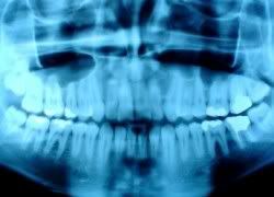
  • Panoramic Dental X-rays show the entire mouth area -- all the teeth in both the upper and lower jaws -- on a single X-ray. This type of Dental X-ray is useful for detecting the position of fully emerged as well as emerging teeth, can identify impacted teeth, and aid in the diagnosis of tumors.
  • Tomograms show a particular layer or "slice" of the mouth while blurring out all other layers. This type of Dental X-ray is useful for examining structures that are difficult to clearly see -- for instance, because other structures are in very close proximity to the structure to be viewed.
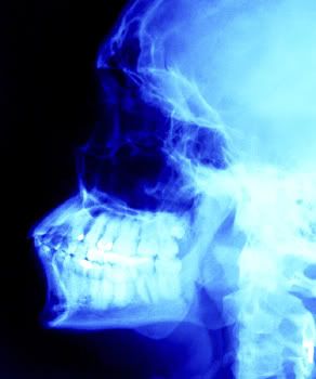
  • Cephalometric projections show the entire side of the head. This type of Dental X-ray is useful for examining the teeth in relation to the jaw and profile of the individual. Orthodontists use this type of X-ray to develop their treatment plans.
  • Sialography involves visualization of the salivary glands following the injection of a dye. The dye, called a radiopaque contrast agent, is injected into the salivary glands so that the organ can be seen on the Dental X-ray film (the organ is a soft tissue that would not otherwise be seen with an X-ray). Dentists might order this type of test to look for salivary gland problems, such as blockages or Sjögren's syndrome.
  • Computed tomography, otherwise known as CT scanning, shows the body's interior structures as a three-dimensional image. This type of Dental X-ray, which may be performed in a hospital or radiology center rather than a dentist's office, is used to identify problems in the bones of the face, such as tumors or fractures. CT scans are also used to evaluate bone for the placement of dental implants and difficult extractions. This helps the surgeon avoid possible complications during and after a surgical procedure.

In the Pipeline

There's a newer dental X-ray technique that your dentist already may be using or may soon be using. It's called digital imaging. Instead of developing X-ray film in a dark room, the X-rays are sent directly to a computer and can be viewed on screen, stored, or printed out. There are several nice benefits of using this new technology including:
  • The technique uses less radiation than the typical X-ray and there is no wait time for the X-rays to develop -- the images are available on screen a few seconds after being taken.
  • The image taken, of a tooth for example, can be enhanced and enlarged many times it's actual size on the computer screen, making it easier for your dentist to show you where and what the problem is.
  • If necessary, images can be electronically sent to another dentist or specialist -- for instance, for a second opinion on a dental problem -- to determine if a specialist is needed, or to a new dentist (if you move).
  • Software added to the computer can help dentists digitally compare current images to previous ones in a process called subtraction radiography. Using this technique, everything that is the same between two images is "subtracted out" from the image leaving a clear image of only the portion that is different. This helps dentists easily see the tiniest changes that may not have been noticed by the naked eye.
Source: WebMD (http://www.webmd.com/oral-health/guide/dental-x-rays-when-get-them)

Dental X-rays May or May NOT Related to Thyroid Cancer?

This is written by a a Filipino writer named Val. You can check this link later to see his information. Just look at the bottom side. Well, I am not really interested about him, what brought me to his link was his article "Dental X-rays is NOT Related to Thyroid Cancer".

Please continue reading...

Dr. Mohammed Al-Samnak, a radiology doctor situated in Kuwait, recently refused on his interview in Kuwait Times that dental x-rays it not related to thyroid cancer. “[The] dental x-ray procedure is very selective or specific. Is a doctor advised only one tooth for an x-ray, our x-ray machine would select only one tooth. It’s not connected to the thyroid, not at all,” Dr. Samnak added.

He stated this due to a new study conducted in Kuwait by scientists from Britain discovered that the exposure to dental x-rays and thyroid cancer is somehow related. Based on this study, patients who were exposed to dental x-rays are ten times more possible to develop thyroid cancer, but this was countered by Samnak. He affirmed confidently that the findings of the study were speculative and unproven.

Dental Xrays

In performing a dental x-ray procedure at any dental clinic in Kuwait, a neck shield is required to use to keep the patient’s thyroid from being exposed to radiation.

Dr. Samnak even told that thyroid cannot possibly acquire thyroid cancer for one or two accidental x-ray exposure. Even though Samnak admittedly stated that thyroid is one of the very sensitive organs in the human’s body, he said that this won’t be a problem due to a neck protection is used during the procedure.

Samnak’s statement was supported by another dentist from Kuwait, Dr Chi Umandap. Dr. Umandap is connected to the office of the assistant undersecretary at the Ministry of Health. According to Umandap, they have used X-ray to pregnant women, when it is for emergency purpose only, and it show no signs of harm done to the baby of the mother.

“Take note though that a pregnant woman should not be exposed to an x-ray, especially during the first three months. In a dental procedure we allow x-rays because they are very safe. Also, radiation dosage is minimal compared to other types of body x-rays,” Dr. Umandap added.

Umandap even explained that a normal x-ray is different to a dental x-ray. In dental-xray, they only used a small dosage of radiation due to only an alveolar bone and tissue cover the teeath.

In contrast to this matter, another doctor, who spoke with Kuwait Times. He countered Dr. Samnak and Dr. Umandap’s ideas. According to this doctor, the findings of this study may possibly accurate. The reason for this is that the dental procedures in Kuwait are very specific, even for a simple tooth ache.

In Kuwait, once a patient complains a dental problem, they will automatically undergo an x-ray, in fact, even schools do such practice. Example, when a tooth is x-rayed first. Then after its removal, another x-ray is advised. Obviously, the patient will be too much exposed to radiation.

The same doctor also stated that in other countries they check the teeth’s condition first before advising them to undergo in an x-ray, if it is necessary. They will ask first the patient’s history then decided whether an x-ray is required or not. Dental x-rays is only done for major orthodontic-cases, the doctor added. Simple dental problem will never require an X-ray.

However, the doctor pointed out that even though government clinics recommend x-ray even for simple tooth ache, repetitive X-raying is never followed by private dental clinics in Kuwait.


Here's another one, I found this in this weblink, written by Mary Shomon... Please continue reading....

Thyroid Cancer Risk May Increase Due to Multiple Dental X-Rays


According to research conducted by UK and Kuwaiti investigators, and recently reported on in the journal Acta Oncologica, the risk of thyroid cancer increases with multiple exposure to dental x-rays.

The thyroid gland is sensitive to radiation, and radiation exposure is a known cause of thyroid cancer. But dental x-rays have long been overlooked as a source of radiation, given the low dose of the radiation used.

Dental Xrays

Repeated exposure, however, now appears to be correlated to an increased risk of thyroid cancer, and according to the researchers, their findings correlate to previous research that has found an increased risk of thyroid cancer in dentists, dental assistants, and x-ray workers.

The researchers found that study subjects who had up to four dental X-rays had more than double the risk of developing thyroid cancer than those who had never had a dental x-ray. Those who had between five and nine X-rays had a risk more than four times normal. And the greatest risk was for those with ten or more X-rays, whose risk was more than five times that of someone who had not received dental x-rays.

Lead investigator Dr. Anjum Memon has said that the implications of the findings are especially important, given the increased rate of thyroid cancer in the past 30 years. In the US, for example, thyroid cancer is the fastest growing form of cancer, and in the United Kingdom, the thyroid cancer rate has doubled from 1.4 per 100,000 in 1975, to 2.9 per 100,000 in 2006.

The researchers are cautioning, however, that the topic requires further research and study, especially to get a better estimate of the radiation doses and number of exposures that are more associated with an increased risk.

What Can You Do?

Given this new information, what should you do to help protect yourself against thyroid cancer?

According to the study author Dr. Memon: "Our study highlights the concern that, like chest or other upper body X-rays, dental X-rays should be prescribed when the patient has a specific clinical need, and not as part of routine check-up or when registering with a dentist. 'So, one of the first things you can do is to make sure that you get dental x-rays only when your dentist has a specific need for them, and not just as a routine part of treatment, i.e., routine annual x-rays, or a routine dental x-ray with every checkup.

Second, and this is a recommendation that I've been making for years here at the site and in my books: Ask the dentist to protect you with a lead thyroid collar when doing any x-rays. This is a recommendation that you'll also hear from About.com's guide to Dentistry, Shawn Watson, who says: "Before you have x-rays taken at your dental office, make sure the protective lead vest, that is placed over your body prior to taking an x-ray, has a thyroid collar." (My own dentist didn't even have a thyroid collar for the longest time. He used to look at me a bit strangely when I would ask that he protect my thyroid for dental x-rays. I would end up pulling the lead apron up so that it would go right up to my chin. Now he has an actual thyroid shield on his lead apron, thankfully!)

Third, if you have young children, minimize their exposure to any unnecessary dental x-rays, and insist that their dentists and orthodontists also use a thyroid collar. Children are especially susceptible to thyroid-damaging radiation, so you'll want to ensure they are not subjected to routine or unneeded dental x-rays. And for children, any essential x-rays should only be conducted with use of the appropriate lead thyroid collar.


I think majority of the dentist did know what are the effects of Dental Xrays if done with too much exposure. In case of ignorance, any dental patient should be aware of some consequences. Dental xrays is good so that the dentist can perform or plan whatever is necessary prior doing any procedure. I believe Dental Xrays are not invented to create such disease but precaution must be undertaken to avoid it.


Source: http://whatisthetrend.net

Related Posts with Thumbnails